SFOV Gamma Camera
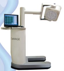
CapIMAGE™ PROVIDES VERSATILITY FOR MULTIPLE PATIENT POSITIONS TO ACCOMMODATE PLANAR, GATED AND DYNAMIC IMAGES
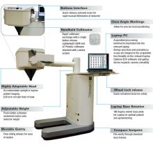
System’s Features- A Number of Options
CapIMAGE is a small field of view gamma camera system ideal for imaging small organs and body parts including the thyroid, parathyroid, planar bone images, and gated cardiac studies. Additionally, CapIMAGE provides a convenient platform for pediatric imaging. CapIMAGE can improve instrumentation and staff productivity by providing an alternative for imaging small organs; thus, freeing up the large field of view gamma cameras for the more complex procedures such as whole body imaging, SPECT and SPECT/CT. This optimization of instrumentation can improve both patient flow as well as staff utilization.
IF DIVERSITY AND QUALITY ARE KEY TO YOUR PRACTICE
CapIMAGE Offers:
- Unique detector stability
- High quality spatial and energy resolution
- Algorhythms optimized for photon position
User-Defined Processing Protocols
- Image filtration
- Flexible display formats
User Defined Acquisition Protocols
- User defined static, dynamic and gated acquisition protocols
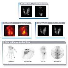
- Thyroid uptakes and imaging
- Parathyroid imaging
- Gated cardiac imaging
- Planar skeletal imaging
DICOM Interface
- Rapid data communication with HIS/RIS and PACS systems
- Worklist data can populate the data fields for the required exam
- Processed studies pushed to the DICOM server for review on any functional workstation
- Optional workstation software available
CREATED FOR USE IN MULTIPLE CLINICAL ENVIRONMENTS AND FOR VARIOUS IMAGING APPLICATIONS
Acquisition and Processing
Hardware: Laptop- Min. 1200 MHz Pentium III Windows Vista
Acquisition functionality:
General:
Persistence scope: 256x256 matrix plus energy spectrum
Pixel size: 4 mm square (64 matrix)
Zoom factors: 1-5 user slectable in 0.1 increments
Image orientation: 0, 90, 180 and 270 degrees
User definable acquisition protocols: Pre-defined acquisitions with all parameters set:
Select acquisition, position camera (manually), hit start.
Manual definition of acquisition protocols
Patient data entry and manipulation: Manual entry (and editing) of patient information
Patient database (via Access and Interfile format)
Patient dataset export: DICOM 3.0. Manual “push and automatic “push” protocol. Configurable (Password protected)
Image display for all dataset types: Selectable gray and color maps / Cine display for dynamics and multi-gated
Static:
Matrix: 64,128, 256, 512 pixels
Energy windows: 3 datasets can be acquired simultaneously
Up to 3 windows can be summed within each
acquired dataset
Total of 9 individual energy regions can be defined
Energy regions cannot overlap
Each window is definied by a centroid energy and a
percent width
Termination:
Automatic stop: Time or counts
Time: 1-10,000 seconds
Counts: 1-1,000,000 kcounts
Camera QC (user accessible):
Uniformity correction Isotope and collimator specific / Intrinsic or extrinsic calibration
Detector peaking: Automated calibration procedure
Uniformity check: Isotope and collimator specific / Intrinsic or extrinsic flood check
“Low count” for visual checks / “High count” for automatic NEMA analysis and reporting / Reporting comprises measured energy peak,
uniformity results and energy resolution
Resolution check: 4 field bar phantom image visual inspection
Camera QC (password protected - Service use):
Detector calibrations: Automated procedures for:
High voltage calibration
PMT tuning
Spatial linearity calibration
Energy linearity calibration
Energy calibration
Geometric calibration
System Specifications
Specifications / CFOV / UFOV
Intrinsic Spatial Resolution
FWHM / <3.7mm / <3.7
FWTM / <7.6mm / <7.6mm
Intrinsic Energy Resolution
FWHM / - / <9.42
Intrinsic Flood Field Uniformity
Integral / <2.5 / <2.5
Differential / <1.5 / <1.5
Intrinisic Spatial Linearity
Absolute / <0.5mm / <0.5mm
Differential / <0.2mm / <0.2mm
Intrinsic High County Rate Performance
Maximum Count Rate 250kps
Environmental Specifications
Temperature/Humidity 15-30° C(59-86° F)
Requirements 20%-80% RH at 25° C(77° F)
Voltage/Current 90-264 VAC, 200VA
Optional Accessories
Diverging Collimator
LEHR Collimator
General
System type: Mobile, single detector system dedicated for planar imaging
Acquisition types: Static, Dynamic, Gated Planar.
Approx. Dimensions 60 cm (W) x 118/173cm (H) x 13-0 cm (D)
23.6” (W) x 46.5/68.1” (H) x 51.2 (D)
Weight: <200 kg (440 lbs.)
Detector Reach >64 cm/25.2” from edge of gantry to center of detector UFOV
Detector Height Approx. 75-130 cm/29.5”-51.2” (parallel hold collimator surface above floor level)
Motions
Detector vertical: Motorized with two speeds
Fast speed: >4.5 cm/sec
Slow Speed: <1 cm/sec
Detector tilt: Manual with lock
Range: >-35 to +90 degrees
Detector Rotate: Manual with Lock
Range: >-90 to +180 degrees


